- Analyzers
- Optics & Sources
- Technologies
- Support
- About
Environmental Scanning Electron Microscopy (ESEM)
In conventional scanning electron microscopy (SEM) systems, the surface of a solid sample is excited with a highly-focused energetic beam of electrons which induces X-ray fluorescence from the elements within the sample. Samples must be run under high vacuum conditions and must be conductive or coated with a conducting material such as gold for proper analysis. X-ray microanalysis of uncoated non-conductive samples in the conventional SEM is hampered by specimen charging which can reduce the energy of the electron beam and make quantitative analysis impossible. Environmental scanning electron microscopy (ESEM) is a unique system in which uncoated biological and industrial materials can be examined with an electron beam in a high chamber pressure atmosphere of water vapor. Therefore, specimens can be analyzed using ESEM without destruction and additional specimen preparation procedures. Dynamic experiments are also possible in the ESEM, such as drying or crystallization.
The main disadvantage of typical ESEM instrumentation is that the electron beam spreads in the high-pressure environmental chamber and excites fluorescent X-rays from the entire specimen, not just from under the electron beam. The fluorescent X-rays generated outside the area of interest are detected by the detector and reduce the image contrast.
A polycapillary optic can be incorporated into an existing ESEM to collect X-rays from an area defined by the optic input focal spot size and focus them on the detector, therefore reducing the background and enhancing the image contrast.
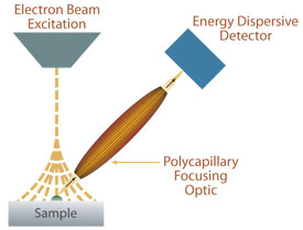
A polycapillary focusing optic as a spatial filter in an ESEM. The XRF signal from a small region within the spread of the electron beam can be selected with the focusing optic.