- Analyzers
- Optics & Sources
- Technologies
- Support
- About
X-ray Optics & Sources
Comprehensive X-ray Source and Optic Solutions
For more than 20 years, XOS has been providing high-quality polycapillary and DCC X-ray optics to both academic and industrial customers worldwide. Our experts work closely with customers to optimize instrument design and performance for a variety of challenging and pioneering applications, on earth and beyond.
Applications
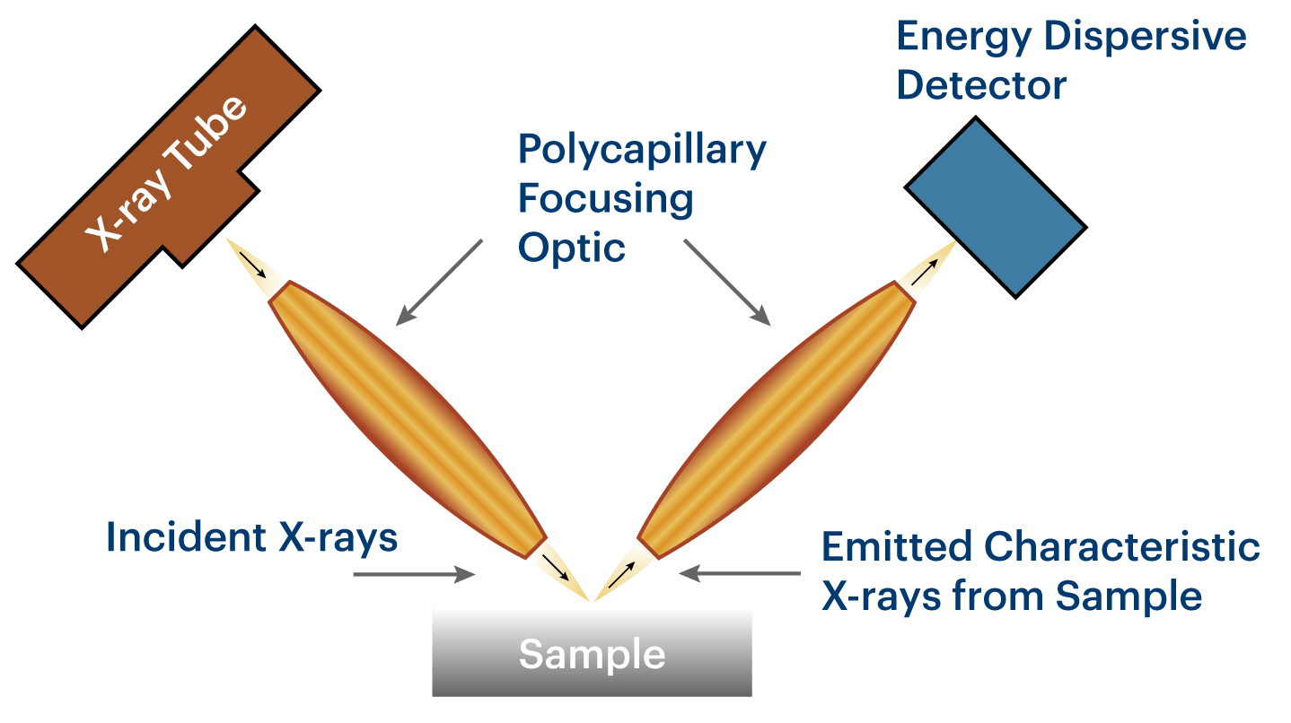
Micro X-ray Fluorescence (μXRF)
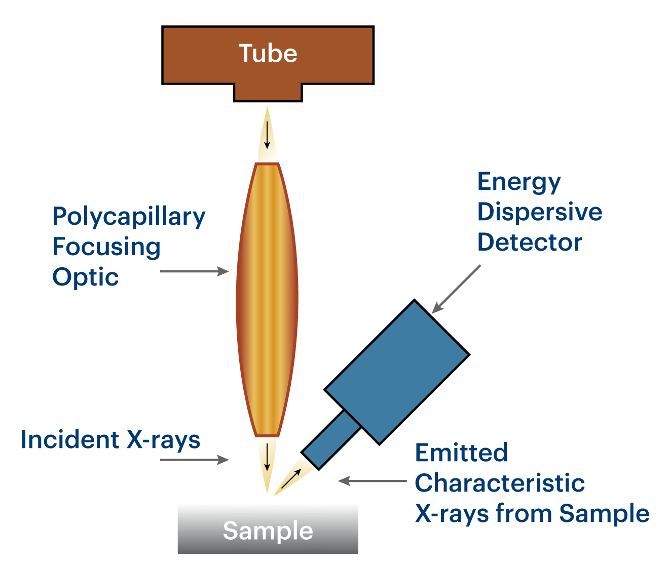
Micro X-ray fluorescence is an elemental analysis technique which allows for the examination of very small sample areas. Like conventional XRF instrumentation, Micro X-ray Fluorescence uses direct X-ray excitation to induce characteristic X-ray fluorescence emission from the sample for elemental analysis. Unlike conventional XRF, which has a typical spatial resolution ranging in diameter from several hundred micrometers up to several millimeters, μXRF uses X-ray optics to restrict the excitation beam size or focus the excitation beam to a small spot on the sample surface so that small features on the sample can be analyzed.
Standard μXRF configuration (on right): The sample can be scanned to measure the elemental distribution within a sample with a spatial resolution as small as 10 μm (energy dependent).
Micro XRF Optic & Source Solutions
X-ray Diffraction (XRD)
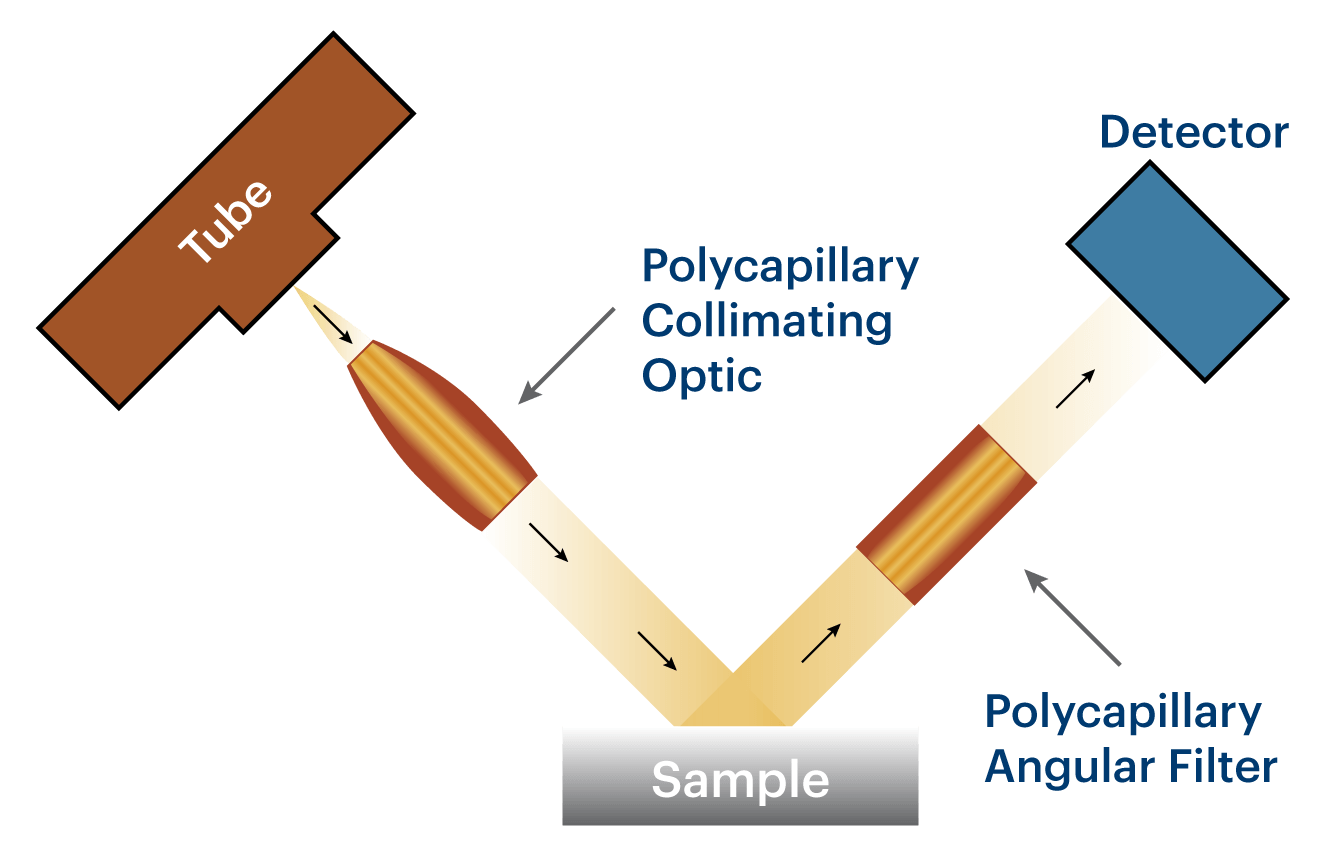
Polycapillary optic can be used in X-ray diffraction (XRD) analysis in two popular configurations, a collimating optic for parallel-beam XRD or a focusing optic for convergent-beam XRD. In parallel-beam XRD (upper figure), the use of a collimating optic can significantly improve the system efficiency so that the performance achieved with a 50-watts X-ray tube is equivalent to that of a conventional system equipped with a kilowatts x-ray tube. Furthermore, the parallel-beam XRD approach makes the system much less sensitive to errors associated with sample irregularity and displacement, eliminating instrument error functions that contribute to peak shift and asymmetric peak broadening.
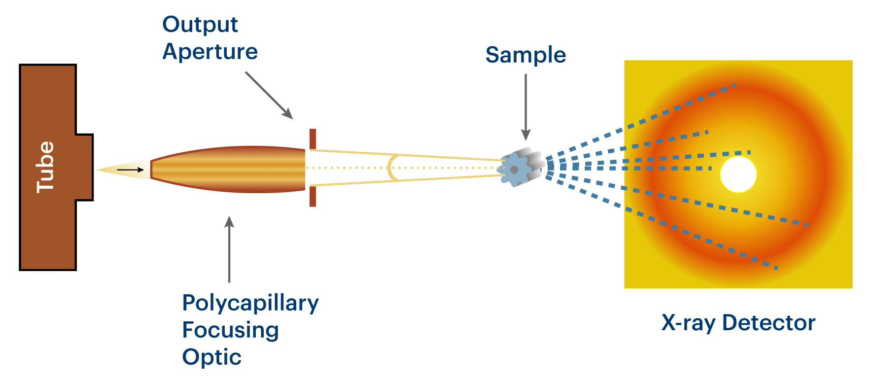
Compared to the parallel-beam XRD applications, convergent-beam XRD systems (lower figure) using polycapillary focusing optics give users more options in achieving small excitation x-ray beam sizes while having the flexibility to optimize the convergent angle and x-ray intensity. The convergent angle of the output beam can be controlled by exchangeable output beam apertures (as shown in the lower figure), a unique feature that allows optimal data quality to be achieve based on sample properties.
Enabled by polycapillary optics, both parallel-beam XRD and convergent-beam XRD analysis have been successfully used in a variety of applications such as phase distribution measurements in the pharmaceutical and steel industries.
- Thin film texture measurements for superconductor layers and magnetic films
- Structure measurements for proteins
- Stress analysis in mechanical components
- Laue X-ray camera and power diffraction for material research
XRD Optic & Source Solutions
Confocal X-Ray Fluorescence (XRF)
The confocal geometry uses two polycapillary focusing optics for enhanced applications of XRF elemental analysis. An excitation optic focuses a small X-ray beam onto the specimen. A detection optic collects fluorescent X-rays from the sample. Specifically, elemental concentrations are measured within the small probe volume (“confocal volume”) defined by the intersection of the output focal spot of the excitation optic and the input focal spot of the collection optic.
This method can be used to detect and quantify elements within radioactive materials. The polycapillary focusing optics act as spatial filters to eliminate background radiation from the sample and increase detection sensitivity for sample elements of interest.
Confocal XRF Optic & Source Solutionos
Parallel-beam Wavelength-Dispersive Spectrometer (WDS)
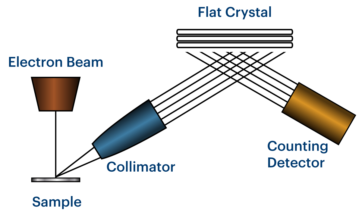
Wavelength-dispersive spectrometers are often coupled in scanning-electron microscope systems. A conventional WDS uses multiple curved crystal monochromators in a complexed configuration to cover the energy range needed. It is bulky, expensive, and difficult to maintain.
In contrast, a modern WDS employs a polycapillary collimating optic to realize the parallel-beam WDS approach, which makes the system simpler, more compact, and more versatile. The use of polycapillary optics allows the fluorescent x-rays from the sample to be converted into a parallel beam that are then monochromatized by multiple flat crystals mounted on a rotating drum.
The parallel-beam approach significantly simplifies the system design and allows the monochromators to be placed at a distance for easy alignment and integration. While PB-WDS devices are only commercially available in configurations to be coupled with SEM systems, custom designed systems have been reported for synchrotron applications.
Parallel-beam WDS Optic & Source Solutions
Synchrotron
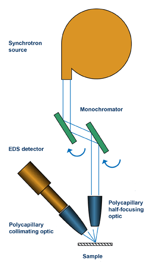
The use of polycapillary focusing optics on synchrotron beam lines are typically in the following two configurations.
-
Half-focusing optic on the excitation beam side: A half-focusing optic guides the nearly parallel incident x-rays, typically off the monochromator assembly, and focuses them to a spot ranging from 5 to 100 microns for micro-X-ray analysis including x-ray fluorescence and x-ray absorption.
While the spatial resolution achieved with a polycapillary optic is nowhere near what a modern x-ray optic can produce (tens of nanometers), it is a popular, cost-effective, and ease-to-use x-ray optical tool for suitable applications. Today, you will find such polycapillary optics in nearly every synchrotron facility around the world.
-
Collimating optic on the detector side: A collimating optic can be used on the detector side in confocal XRF configurations or as a spatial filter device to reduce the background generated by the scattered x-rays. The excitation beam can be generated by a variety of x-ray optics including a polycapillary focusing optic.
Synchrotron Optic & Source Solutions

Products
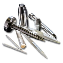
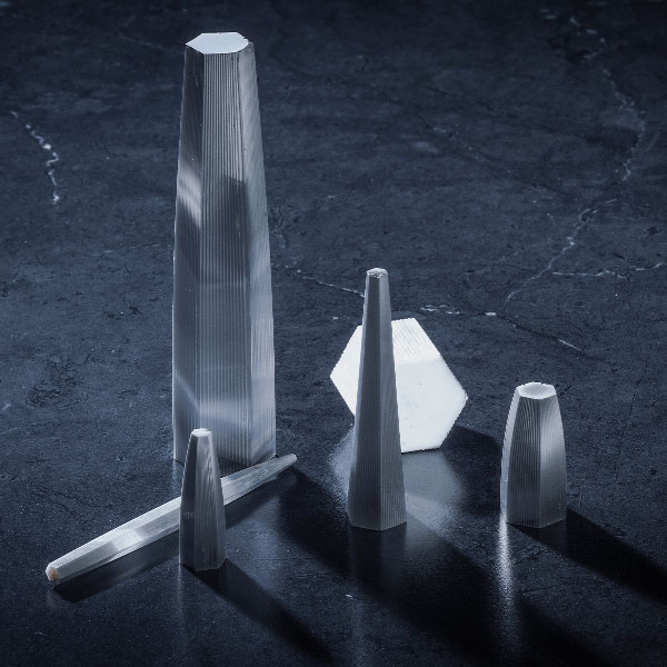
Polycapillary Optics
XOS Polycapillary Optics capture large, solid angles of X-rays and redirect them to a micron-sized focal spot or a highly collimated beam.
Features & Benefits
- Significantly improved measurement speed for fine-feature analysis and high-resolution mapping
- Halo reduction optics optimized for high-energy applications
- Large, quasi-parallel X-ray beam for XRD and XRF
- Customized optic design for optimal performance
Applications
- Micro-XRF for elemental mapping, plating thickness, and fine feature analysis
- Confocal XRF analysis and wavelength-dispersive spectrometer (WDS)
- Synchrotron-based micro-X-ray analysis (Micro-XRF, micro-XAS and confocal XRF)
- Powder XRD, texture and stress analysis
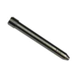
Monocapillary Optics
XOS single-bounce monocapillary optics can convert incident parallel X-rays to a micron-sized focal spot by total reflections.
Features & Benefits
- Micron-sized focal spot with large working distance
- Flux density gain up to three orders of magnitude
- Compact and easy mount
- Broadband energy range
- Custom design and solutions
Applications
- Synchrotron beamline applications
- Large distance, incident parallel X-rays
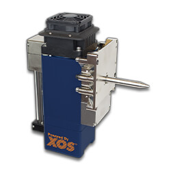
fleX-Beam
The XOS fleX-Beam is a compact X-ray generator combining a low-powered X-ray source and a precisely aligned polycapillary optic.
Features & Benefits
- Intensity is up to 10,000 times greater than conventional pinhole collimators
- Focal spot as small as 5μm @ Rh Ka (20.162keV)
- 50-watt performance exceeds conventional kilowatt-powered X-ray tubes
- Integrated safety shutter & 8-position filter wheel
- Compact; easily integrates with any instrument or system
- Interchange different optics, as well as service the X-ray source in the field
Applications
- Micro XRF for small feature analysis, film and plating thickness, high-resolution elemental mapping
- XRD for residual stress analysis
- Laue Diffraction
- Powder Diffraction
- XRD & WDS
- Powder Diffraction
- Texture & Strain Measurement
- Wavelength-Dispersive Spectrometer
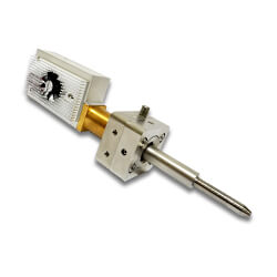
Mini-Beam
The XOS Mini-Beam is a compact and powerful X-ray generator that integrates a polycapillary X-ray optic with a low powered X-ray source.
Features & Benefits
- Mini-Beam’s intensity is up to 1,000 times greater than conventional pinhole collimators
- Focal spot as small as 5μm @ Rh Ka (20.16keV)
- 50-watt performance exceeds conventional kilowatt-powered X-ray tubes
- Integrated removable filter for simple swapping
- Compact geometry for applications with limited space or portability requirements
- User friendly fine optic alignment mechanism and simplistic mount for optic and/or source swapping
Applications
- Micro X-ray fluorescence (micro-XRF)
- Small Feature Analysis Film & Plating Thickness
- High-Resolution Elemental Mapping
- Gun residue study in forensics
- Pigment study in art preservation
- Composition and artifact analysis in archaeology
- X-ray diffraction (XRD)
- Residual Stress Analysis
- Laue Diffraction
- Powder Diffraction
Contact Us
Our industry experts are here to help you create the perfect custom solution for your application needs.